您的位置:首页 > 产品中心 > RIPAb+ hnRNP U-RIP Validated Antibody and Primer Set
RIPAb+ hnRNP U-RIP Validated Antibody and Primer Set
产品别名
RIPAb+ hnRNP U-RIP Validated Antibody and Primer Set
Scaffold attachment factor A, heterogeneous nuclear ribonucleoprotein U, heterogeneous nuclear ribonucleoprotein U (scaffold attachment factor A), hnRNP U, hnRNP U protein, p120 nuclear protein
基本信息
| eCl@ss | 32160702 |
| NACRES | NA.32 |
| General description【一般描述】 | RIPAb+ antibodies are evaluated using the RNA Binding Protein Immunoprecipitation (RIP) assay. Each RIPAb+ antibody set includes a negative control antibody to ensure specificity of the RIP reaction and is verified for the co-immunoprecipitation of RNA associated specifically with the immunoprecipitated RNA binding protein of interest. Where appropriate, the RIPAb+ set also includes quantitative RT-PCR control primers (RIP Primers) to biologically validate your IP results by successfully co-precipitating the specific RNA targets, such as messenger RNAs. The qPCR protocol and primer sequences are provided, allowing researchers to validate RIP protocols when using the antibody in their experimental context. If a target specific assay is not provided, the RIPAb+ kit is validated using an automated microfluidics-based assay by enrichment of detectable RNA over control immunoprecipitation. Proteins of the heterogeneous nuclear ribonucleoparticles (hnRNP) family form a structurally diverse group of RNA binding proteins implicated in various functions. Recently, hnRNP proteins have been shown to hinder communication between factors bound to different splice sites. hnRNP-U, also termed scaffold attachment factor A (SAF-A), binds to pre-mRNA and nuclear matrix/scaffold attachment region DNA elements. |
| Specificity【特异性】 | This antibody recognizes hnRNP U. |
| Immunogen【免疫原】 | Recombinant protein corresponding to human hnRNP U. Epitope: Unknown |
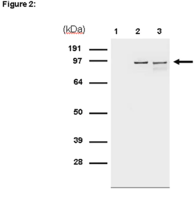
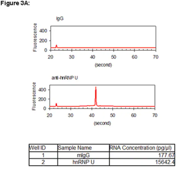
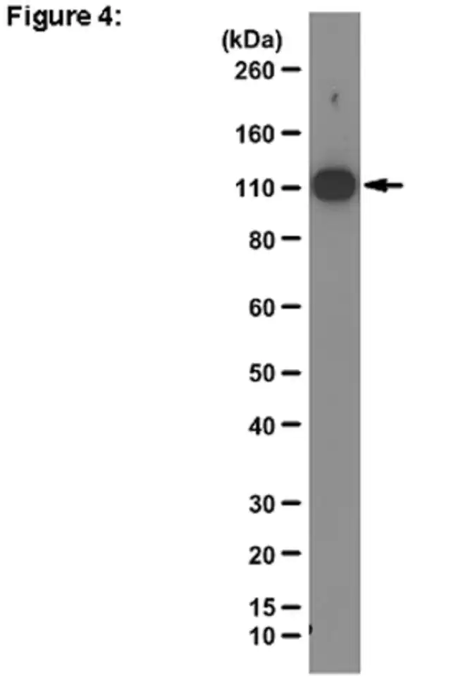
| Application【应用】 | Immunoprecipitation from RIP lysate: Representative lot data. RIP lysate from HeLa cells (~2 X 10E7 cell equivalents per IP) was subjected to immunoprecipitation using 5 µg of either a normal mouse IgG, (Cat. # CS200621), or 5 µg of Anti-hnRNP U antibody (Cat. # CS207320). ten percent of the precipitated proteins (lane 1: normal mouse IgG, lane 2: hnRNP U) and HeLa whole cell lysate (lane 3) were resolved by electrophoresis, transferred to nitrocellulose and probed with anti-hnRNP U antibody (Cat. # CS207320, 1:1000). Proteins were visualized using One-Step Arrow indicates hnRNP U. (Figure 2). Automated Microfluidics-based Electrophoretic RNA Separation and Analysis (MFE): Representative lot data. RIP Lysate prepared from HeLa cells (2 X 10E7 cell equivalents per IP) were subjected to immunoprecipitation using 5 µg of either 1. normal mouse IgG (Cat. # CS200621), or 2. Anti-hnRNP U antibody (Cat. # CS207320) and the Magna RIP Successful immunoprecipitation of hnRNP U-associated RNA was verified by automated microfluidics-based electrophoretic RNA separation and analysis. Please refer to the Magna RIP Western Blot Analysis: Representative lot data. K562 cell lysate was probed with Anti-hnRNP U, clone 3G6 (0.01 µg/mL). Proteins were visualized using a Goat Anti-Mouse IgG secondary antibody conjugated to HRP and a chemiluminescence detection system. Arrow indicates hnRNP U (~120 kDa). (Figure 4). Research Category Epigenetics & Nuclear Function Research Sub Category RNA Metabolism & Binding Proteins Apoptosis - Additional This RIPAb+ hnRNP U -RIP Validated Antibody & Primer Set conveniently includes the hnRNP U antibody & the specific control PCR primers. |
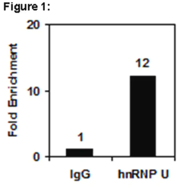
| Quality【质量】 | RNA Binding Protein Immunoprecipitation: Representative lot data. RIP Lysate prepared from HeLa cells (2 X 10E7 cell equivalents per IP) were subjected to immunoprecipitation using 5 µg of either a normal mouse IgG or 5 µg of Anti-hnRNP U antibody and the Magna RIP® RNA-Binding Protein Immunoprecipitation Kit (Cat. # 17-700). Successful immunoprecipitation of hnRNP U-associated RNA was verified by qPCR using RIP Primers Ribosomal Protein S19, (Figure 1). Please refer to the Magna RIP |
| Physical form【外形】 | Format: Purified Anti-hnRNP U (Mouse Monoclonal), Part # CS207320. One vial containing 50 µg of protein G purified mouse IgG1 in 0.1M Tris-glycine, pH 7.4, 0.15M NaCl, 0.05% sodium azide and 30% glycerol. Store at -20°C. Normal Mouse IgG, Part # CS200621. One vial containing 125 µg of purified mouse IgG in 125 µL of storage buffer containing 0.1% sodium azide. Store at -20°C. RIP Primers, Ribosomal Protein S19, Part # CS207321. One vial containing 75 μL of 5 μM of each primer specific for human c-myc 3′ UTR. Store at -20°C. FOR: ACG CGA GCT GCT TCC ACA G REV: AGC TGC CAC CTG TCC GGC Protein G Purified |
| Analysis Note【分析说明】 | Control Includes negative control normal mouse IgG antibody and control primers specific for the cDNA of human Ribosomal Protein S19. |
| Other Notes【其他说明】 | Concentration: Please refer to the Certificate of Analysis for the lot-specific concentration. |
| Legal Information【法律信息】 | MAGNA RIP is a registered trademark of Merck KGaA, Darmstadt, Germany UPSTATE is a registered trademark of Merck KGaA, Darmstadt, Germany |
产品性质
| Quality Level【质量水平】 | 100 |
| biological source【生物来源】 | mouse |
| antibody form【抗体形式】 | purified immunoglobulin |
| clone【克隆】 | 3G6, monoclonal |
| species reactivity | human |
| manufacturer/tradename | RIPAb+ Upstate® |
| technique(s) | RIP: suitable western blot: suitable |
| isotype【同位素/亚型】 | IgG2bκ |
| NCBI accession no.【NCBI登记号】 | NP_004492.2 |
| UniProt accession no.【UniProt登记号】 | Q00839 |
| shipped in【运输】 | dry ice |
| packaging【包装】 | 10 assays per set. Recommended use: ~5 μg of antibody per RIP (dependent upon biological context). |
产品说明
| Target description【目标描述】 | The calculated molecular weight is 90 kDa However, the protein is usually observed at ~120 kDa (Dreyfuss, G., et al. (2002). Nat Rev Mol Cell Biol. 3(3):195-205.) |
| Storage and Stability【储存及稳定性】 | Stable for 1 year at -20°C from date of receipt. Handling Recommendations: Upon receipt, and prior to removing the cap, centrifuge the vial and gently mix the solution. Aliquot into microcentrifuge tubes and store at -20°C. Avoid repeated freeze/thaw cycles, which may damage IgG and affect product performance. Note: Variabillity in freezer temperatures below -20°C may cause glycerol containing solutions to become frozen during storage. |
| Disclaimer【免责声明】 | Unless otherwise stated in our catalog or other company documentation accompanying the product(s), our products are intended for research use only and are not to be used for any other purpose, which includes but is not limited to, unauthorized commercial uses, in vitro diagnostic uses, ex vivo or in vivo therapeutic uses or any type of consumption or application to humans or animals. |
安全信息
| Storage Class Code【储存分类代码】 | 10 - Combustible liquids |

